

Flashes and Floaters
Understanding posterior vitreous detachment and retinal tears
Flashes and Floaters

Have you recently developed new flashes or floaters?
Sudden onset flashes, a shower of new floaters, a curtain or shadow across your vision, or sudden reduction in vision require urgent ophthalmic assessment. If you are unable to access an ophthalmologist promptly, attend your optometrist or nearest hospital emergency department.
Flashes and floaters are common visual symptoms that can occur when the vitreous gel inside the eye changes with age. In many cases, they are caused by a posterior vitreous detachment (PVD), which is a common age-related separation of the vitreous gel from the retina.
Patients often describe floaters as threads, cobwebs, hair like opacitieis or dots and shadows that move with the eye. They are usually most noticeable when looking at a plain bright background, such as a blue sky, white wall or computer screen. Flashes are often seen as brief arcs or streaks of light in the peripheral vision, commonly in one eye and often more noticeable in the early evening soon after it has become dark and during the night.
Urgent assessment is recommended if flashes or floaters start suddenly, if there is a shower of new floaters, blurred vision, a shadow or curtain in the vision. These symptoms may indicate a retinal tear or retinal detachment, which can cause loss of vision (blindness) if not urgently identified and treated.
What Causes Flashes and Floaters?
The eye is filled with a clear gel called the vitreous. With age, the vitreous gradually becomes more liquid and can separate from the retina. This is called a posterior vitreous detachment or PVD. It is common, particularly from middle age onwards, and is one of the most frequent causes of new floaters and flashes.
Floaters occur when small condensations within the vitreous cast shadows on the retina. A larger ring-shaped floater may occur when the vitreous separates from around the optic nerve. Flashes occur when the vitreous pulls on the retina. The retina interprets this mechanical traction as light, even though no external light is present.
In most people, the vitreous separates without causing damage. In some cases, however, the traction is strong enough to tear the retina. A retinal tear is important because fluid can pass through the break and lift the retina away from the back of the eye, causing a retinal detachment.
%20with%20vitreous%20floater.jpg)
Posterior vitreous detachment with the vitreous separating from the retina and associated floaters
Posterior Vitreous Detachment (PVD)
A posterior vitreous detachment (PVD) is not the same as a retinal detachment. PVD is usually a normal ageing change inside the eye. It is not painful, and in many people the symptoms gradually become less noticeable over time.
The critical issue is whether the vitreous has caused a retinal tear during the process of separation. A thorough dilated retinal examination is required to assess the peripheral retina, where retinal tears often occur.
Floaters from a PVD may persist, but many patients become less aware of them over weeks to months as the floater moves away from the central visual axis or the brain adapts to its presence.
Retinal Tear
A retinal tear can develop when the vitreous pulls firmly on the retina during a posterior vitreous detachment. Most retinal tears occur in the far peripheral retina and may not immediately affect central vision. Patients commonly notice new flashes, a sudden increase in floaters, or occasionally a small amount of bleeding into the vitreous cavity.
The importance of recognising a retinal tear is that it can often be treated before a retinal detachment develops. If identified early, laser retinopexy is usually performed as an outpatient procedure. The laser creates a barrier of scar tissue around the tear, helping to prevent fluid passing beneath the retina. Occasionally cryotherapy may be necessary to seal the tear.
Prompt diagnosis and treatment is needed to reduce the risk of vision-threatening complications.
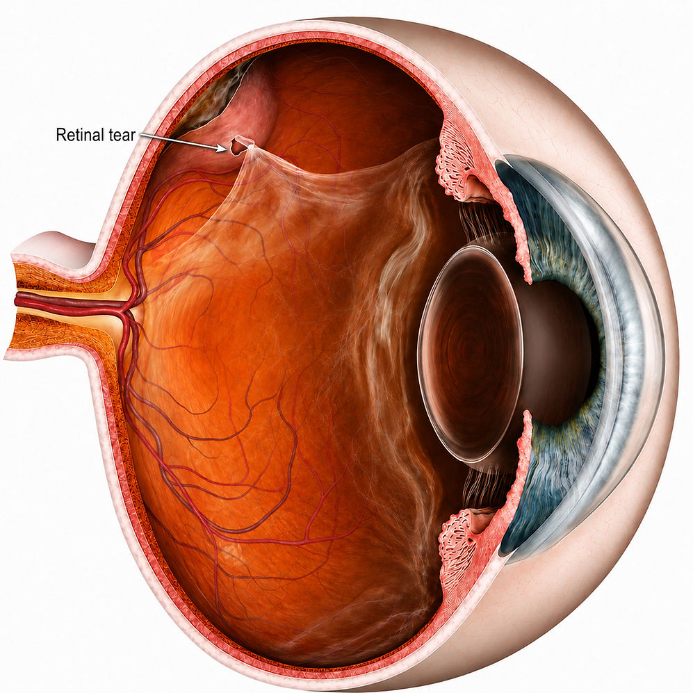
Retinal tear due to vitreous traction
Risk Factors for Retinal Tears
Risk factors are not required to develop a retinal tear, but they make the likelihood greater. These factors include*:
-
Advanced age
-
Degree of myopia (short-sightedness)
-
Associated lattice degeneration (thin patches in the retina) or peripheral retinal degeneration
-
Eye Trauma
-
Family history of retinal tears or detachment
-
Prior eye surgery
There is no definite way to predict who might develop a retinal tear or when it might occur. All patients with new onset floaters, flashes or curtain/shadow in vision are treated with suspicion and require urgent ophthamic evaluation.
* from the American Society of Retina Specialists (ASRS)
Retinal Detachment
A retinal detachment occurs when fluid passes through a retinal tear and separates the retina from the underlying tissue that supplies it with oxygen and nutrients. Unlike a retinal tear, a retinal detachment usually causes progressive loss of vision and requires urgent surgical treatment.
Patients often describe a dark curtain or shadow moving across part of their vision, increasing peripheral vision loss, or sudden blurred vision. The location of the shadow depends on where the detachment begins. If left untreated, the detachment may extend towards the macula, resulting in permanent central vision loss.
A retinal detachment is a sight-threatening condition that usually requires urgent surgery. Although surgery maynsuccessfully reattach the retina, the visual prognosis is frequently guarded and depends on several factors, particularly whether the macula has detached before treatment.
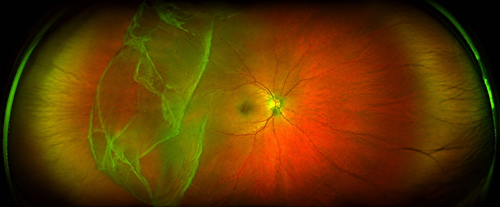
Ultra widefield retinal photograph demonstrating a temporal rhegmatogenous retinal detachment threatening the macula
When Should You Seek Urgent Attention?
You should arrange prompt ophthalmic assessment if you develop new flashes or floaters, particularly if symptoms are sudden or occur in one eye.
Urgent assessment is especially important if there is a sudden shower of floaters, flashes with blurred vision, a dark shadow or curtain, reduced peripheral vision, eye trauma, high myopia, previous retinal tear or detachment, or recent eye surgery.
Assessment involves using dilating drops so the peripheral retina can be examined properly.
Your vision and eye pressure will be checked, and the retina will likely be assessed using slit lamp biomicroscopy, specialised lenses and widefield imaging where appropriate. A binocular indirect ophthalmoscope may be used. Gentle pressure will need to be applied to indent the peripheral retina to ensure no small peripheral tears are undetected.

Examining the retina for retinal tears and checking for retinal detachment
Barrier Laser Retinopexy
The aim of laser retinopexy is to reduce the risk of a retinal tear progressing to a retinal detachment. By sealing the retina around the tear, laser treatment is usually effective in significantly reducing the risk of retinal detachment and can avoid the need for more complex retinal surgery.
Laser retinopexy is generally a safe procedure and is usually performed in the consulting rooms using topical anaesthetic eye drops.
As with any medical procedure, small risks can occur. These may include temporary blurred vision, mild discomfort during treatment, inflammation and a temporary increase in floaters. Occasionally, further laser treatment or surgery may be required. Our ophthalmologists will discuss in greater detail the risks with you and answer your questions. Given the aim is to reduce the serious complication of retinal detachment and loss of vision, barrier laser retinopexy is usually performed urgently as soon as possible once a retinal tear is identified.
Regular follow-up and prompt review of any new flashes, floaters or changes in vision remain important.

Peripheral retinal tear before and during encircling barrier laser retinopexy
At Eye and Retina Specialists Sydney, patients with flashes and floaters undergo careful assessment of the vitreous and retina to exclude retinal tears and retinal detachment. We are located in Green Square, located between the CBD and Sydney Airport with patient parking available. Prompt examination is particularly important when symptoms are new, sudden or changing.
Our practice is equipped for detailed retinal assessment, including dilated examination and retinal imaging where appropriate. If a retinal tear is identified, timely treatment may reduce the risk of progression to retinal detachment.