
Central Serous Retinopathy (CSR) Assessment & Management Sydney | Retina Specialist
Central Serous Retinopathy (CSR), also known as Central Serous Chorioretinopathy (CSCR), is a retinal condition in which fluid accumulates beneath the central retina. It often affects the central part of vision, at the macula. At Eye & Retina Specialists, we provide comprehensive assessment, advanced retinal imaging and management of CSR from our Sydney retina clinic located in Green Square.
The retina is the light-sensitive tissue lining the back of the eye. In CSR, fluid leaks from the choroid, a vascular layer beneath the retina, through the retinal pigment epithelium (RPE) and accumulates beneath the macula. The resulting separation of the neurosensory retina from the underlying RPE can cause visual symptoms ranging from mild blurring to significant distortion.
Most patients experience a single episode that resolves spontaneously, however some develop recurrent or chronic disease requiring ongoing monitoring and treatment.
CSR is increasingly recognised as part of the pachychoroid disease spectrum, a group of retinal conditions characterised by abnormalities of the choroidal circulation and increased choroidal thickness.
What is Central Serous Retinopathy (CSR) ?
Slider demonstrating the difference between a normal macula OCT and an eye with acute CSR
Symptoms of Central Serous Retinopathy
CSR typically appears suddenly and predominantly impacts one eye at a time, though it can occur bilaterally. Symptoms may develop rapidly and can include:
-
A dark, dull, or grey patch directly in your central line of sight.
-
Distortion of straight lines (metamorphopsia), where doorframes or lines of text appear wavy, bent, or kinked.
-
Objects appearing smaller (micropsia) or further away than expected.
-
Difficulty reading and a noticeable reduction in colour perception, contrast, or low-light clarity.
Urgent Retinal Assessment: If you are experiencing sudden central vision distortion or a blind spot, contact our Waterloo clinic on (02) 9699 0001 or email reception@eyeandretina.com.au to schedule a priority consultation with a retina specialist. If sending an email, write "URGENT" in the subject line.

Visual simulation of central serous retinopathy, demonstrating the characteristic central blur and distortion
Causes and Risk Factors for Central Serous Retinopathy
While the precise underlying cause is still being researched, current evidence suggests that abnormalities within the choroid (the vascular layer beneath the retina) result in increased vessel permeability, causing fluid to leak through the Retinal Pigment Epithelium (RPE).
CSR most commonly affects adults between the ages of 30 and 60, appearing up to six times more frequently in men than in women.
Key clinical risk factors include:
Psychological Stress & Personality: High-stress lifestyles or classic "Type A" personality traits significantly increase susceptibility due to elevated systemic cortisol levels.
Corticosteroid Exposure: Steroids are one of the most potent triggers for CSR. This includes prescription oral medications, joint injections, inhaled steroid medications, nasal steroid sprays, and even over-the-counter anti-inflammatory topical steroid creams.
Carotenoid & Antioxidant Depletion: Emerging serum analyses indicate that individuals experiencing an active CSR flare-up often display a lower systemic antioxidant capacity and lower circulating plasma lutein levels, which may compromise the macula's natural defense against oxidative stress.
Systemic Conditions: Obstructive sleep apnoea, uncontrolled high blood pressure (hypertension), and pregnancy can also heighten risks.
Note: In many patients, an episode can occur without an obvious identifiable trigger.

Key clinical risk factors associated with central serous retinopathy (CSR)
How Is CSR Diagnosed?
Accurate staging and tracking of CSR requires a comprehensive retinal examination combined with high-definition structural and vascular imaging to confirm the diagnosis and assess disease activity. At Eye & Retina Specialists, our diagnostic suite includes:
Optical Coherence Tomography (OCT)
OCT provides non-invasive, high-resolution cross-sectional imaging of the retina and is the most critical investigation for diagnosing and monitoring CSR. It allows us to objectively measure retinal thickness, map subretinal fluid beneath the macula, and identify Pigment Epithelial Detachments (PEDs) to precisely track treatment response.
Fluorescein Angiography (FFA)
Performed when the exact source of leakage needs to be mapped for laser planning, an FFA utilizes a safe, biocompatible digital dye to identify active leakage points. This reveals classic, distinct leakage footprints, such as the "ink-blot" or expanding "smokestack" leakage patterns.
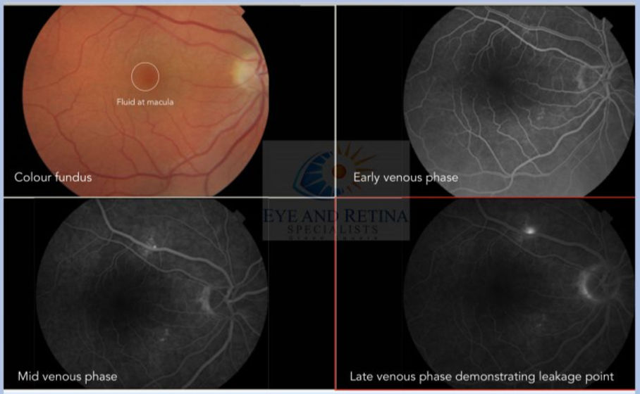
Fluorescein angiography demonstrating a focal leakage point in Central Serous Retinopathy (CSR)
Indocyanine Green Angiography (ICG)
Particularly useful for chronic or recurrent cases, ICG angiography allows our specialists to look deeper into the ocular blood supply, demonstrating underlying choroidal hyperpermeability and structural pachychoroid changes that standard imaging cannot as reliably detect.

Multimodal retinal imaging demonstrating central serous retinopathy with subretinal fluid visible on OCT, fundus imaging and ultrawide field ICG Angiography of the right and left eye
Acute Versus Chronic CSR
The clinical management and long-term visual outcome of Central Serous Retinopathy depend heavily on the duration of the disease process.
Acute CSR is defined as disease duration lasting six months or less. Most cases of acute CSR follow self-limiting path where the subretinal fluid undergoes spontaneous resorption within this timeframe. During this time it is important to treat any underlying potential causes, avoid or reduce corticosteroid use where possible and aim to reduce life stressors if possible.
While the immediate visual prognosis is generally good, acute cases require proactive clinical oversight rather than passive waiting. Patients must remain highly vigilant by tracking visual symptoms closely, as any sudden increases in central blurring, alterations in colour contrast, or the expansion of a central blind spot require immediate review.
To ensure early detection of subtle changes in visual distortion or new areas of central vision loss, patients are instructed to utilize an Amsler Grid daily at home.
Regular OCT imaging is important during this acute monitoring phase. These serial scans allow clinicians to objectively measure the height and volume of the subretinal fluid and monitor the structural health of the underlying photoreceptor layer.
Chronic CSR is formally diagnosed when subretinal fluid and symptoms persist continuously for more than six months.
In chronic presentations, the prolonged presence of fluid can cause progressive, irreversible damage to both the delicate photoreceptors and the underlying Retinal Pigment Epithelium cells. This chronic fluid exposure may lead to localised tissue breakdown, chronic contrast loss, and permanent central vision reduction. For this reason, shifting from conservative observation to active, evidence-based intervention is usually recommended once the six-month threshold is crossed to reduce permanent macular impairment.

Persistent fluid in chronic Central Serous Retinopathy can result in progressive retina damage and long term visual impairment
Management of CSR
Most cases of CSR resolve without treatment, usually within 6 months of onset. During this period it is important that the vision and fluid be monitored with regular checks. Where possible, try to reduce stressors in your life activities. In consultation with your GP and medical team, try to avoid corticosteroid use.
If the CSR does not resolve within 6 months, it is termed “chronic CSR” and requires medical intervention. This is because the risks of visual impairment from persisting subretinal fluid become more significant.
The appropriate treatment is based on the leakage point, as shown by Fundus Fluorescein Angiography (FFA) and ICG Angiography. Our clinic is equipped with ultra-wide field FFA and ICG imaging, allowing both angiograms to be performed within a short period of time using interweaved technology. ICG imaging allows visualisation of the deeper choroidal circulation, which can more clearly delineate the source of leakage. This specialised test is only available at a limited number of ophthalmology centres.
Treatment Options
There is no single treatment option suitable for all cases of CSR, and treatment is usually guided by the location of the leakage point. There are risks and benefits with every treatment option that needs to be considered.
If the leakage point is away from the central vision, retinal laser may be appropriate. Half-fluence photodynamic therapy (PDT) has been shown to be effective for the treatment of CSR, and this may be more appropriate if the leakage point is closer to the central vision. Micropulse laser is another option in this situation.
Our retinal specialists will discuss the various treatment modalities with you in further detail should this be required. The main treatment option can be briefly summarised as follows:
Observation & Monitoring
Best Suited For: First-episode acute cases (under 3–6 months) with minimal visual disruption.
How it Works: Regular monitoring with high-resolution OCT to track natural fluid reabsorption while addressing lifestyle and systemic risk factors. Regular home monitoring with Amsler Grid chart.
Risk Factors & Nutritional Support
Best Suited For: All diagnosed patients, particularly those showing signs of chronicity or recurrence.
How it Works: Safe tapering of steroid medications with your prescribing physician, managing sleep apnea, and optimizing blood pressure. Clinical studies also support initiating targeted oral supplementation (20mg Lutein + Zeaxanthin) to accelerate fluid clearance and protect macular pigment density from oxidative fluid stress.
Photodynamic Therapy (PDT)
Best Suited For: Chronic CSR (fluid persisting past 6 months) for leaks located near the central fovea.
How it Works: A light-sensitive medication (Visudyne) is infused intravenously to accumulate in the leaky choroidal vessels. A cold, non-thermal laser then targets the area, safely closing down the hyper-permeable vessels without scarring the delicate retina.
Subthreshold Retinal Laser Therapy
Best Suited For: Fluid leaks located close to or directly under the center of vision (the fovea).
How it Works: A highly precise laser applies energy at a level below the threshold of cellular damage. This triggers a natural biologic response that stimulates the pumping action of the retinal pigment epithelium (RPE) cells to drain away the subretinal fluid, completely avoiding the destructive heat, thermal tissue damage, or scarring associated with conventional lasers.
Conventional Argon Laser
Best Suited For: Targeted leaks located safely away from the central macular zone.
How it Works: A targeted thermal laser delivers localized heat directly to the pinpoint source of the leak, sealing it shut to halt fluid production immediately.
Oral Medications (Spironolactone / Eplerenone)
Best Suited For: Not routinely recommended. Historically considered for chronic presentations.
How it Works: While oral mineralocorticoid receptor blockers were common past therapies, they have largely fallen out of favor and are no longer standard practice. Major recent international clinical publications (including the landmark VICI trial) demonstrated that these oral systemic medications show no long-term visual or structural benefits over simple observation.

Management options for Central Serous Retinopathy range from observation and monitoring through to laser-based treatments in selected patients
If you have been diagnosed with CSR or are experiencing unexplained visual distortions, early retinal specialist evaluation is important to aim to protect your long-term macular health.
Our medical retinal specialist Dr Neil Sharma continues regular academic, teaching and research interests and operates advanced diagnostic and ultra widefield retinal imaging suite. He is a member of the Australian and New Zealand Society of Retinal Specialists and is a recognised subspeciality expert in custom individualised medical retina and laser interventions.
Phone: (02) 9699 0001
Email: reception@eyeandretina.com.au
Clinic Location: Suite C1, 30 O'Dea Avenue, Waterloo, NSW 2017