Our Equipment
Advanced Ophthalmic Technology and Diagnostic Imaging
Modern ophthalmology relies heavily on precise imaging, detailed diagnostic assessment and longitudinal monitoring of ocular disease.
At Eye and Retina Specialists, we utilise a comprehensive suite of advanced ophthalmic imaging and diagnostic technologies to assist in the assessment, diagnosis and management of retinal disease, cataract, glaucoma, refractive conditions and complex medical eye disorders.
Our practice incorporates contemporary retinal imaging, optical coherence tomography (OCT), ultra-widefield photography, visual field testing, ocular biometry and anterior segment diagnostic systems within an integrated clinical workflow. These technologies assist our ophthalmologists in detecting subtle disease, monitoring progression over time and planning treatment with greater precision.


Comprehensive Ophthalmic Imaging and Diagnostic Assessment
Ophthalmologists undertake many years of medical and surgical training in the diagnosis and management of complex eye disease. Modern ophthalmology increasingly relies on advanced imaging and diagnostic technologies to assist in the optimum management of retinal, macular, optic nerve, corneal and anterior segment disorders. This assists in providing the highest possible patient outcomes and satisfaction.
Our practice utilises a broad range of ophthalmic diagnostic systems in the evaluation of conditions including macular degeneration, diabetic eye disease, retinal vascular disease, epiretinal membrane, vitreomacular traction, glaucoma, optic nerve disorders, hydroxychloroquine retinal toxicity and peripheral retinal pathology. Additional technologies such as ocular biometry, corneal imaging and visual field analysis also assist in cataract surgery planning, refractive assessment, driving vision testing and aviation ophthalmology examinations.
Advanced ophthalmic investigations provide comprehensive structural, vascular and functional information. Our practice has invested in advanced ophthalmic imaging and diagnostic technologies from established global manufacturers including Haag-Streit, Keeler, ZEISS, Optos, Topcon, Ellex and Sonomed Escalon. The availability of these systems within the practice allows multiple investigations to be performed efficiently within the one clinical environment.

High-magnification slit lamp biomicroscopy with LED illumination is fundamental to a detailed examination of the front and back parts of the eye
Integrated Imaging and Longitudinal Analysis
Many ophthalmic conditions require careful comparison of imaging and functional testing over extended periods rather than interpretation of isolated scans alone. Our practice utilises ZEISS FORUM software integration, including Retina Workplace and Glaucoma Workplace platforms, allowing multimodal diagnostic data to be reviewed cohesively within a unified clinical interface.
Each clinical review contributes an additional longitudinal data point, allowing subtle structural and functional changes to be compared across increasingly robust datasets over time. This may assist in identifying progression trends earlier and improving the accuracy of long-term disease monitoring in chronic retinal and glaucoma-related conditions.
By integrating OCT imaging, retinal photography and visual field analysis across multiple visits, these platforms assist with more detailed longitudinal assessment and increasingly individualised management decisions over time.
Because structural and functional progression in retinal and glaucoma disease may occur gradually, longitudinal analysis can be particularly valuable when attempting to distinguish true disease progression from normal test variability or isolated fluctuation. Careful comparison of historical imaging and visual field data may assist in identifying clinically meaningful change earlier and with greater confidence over time.

ZEISS FORUM software integrating longitudinal OCT imaging and retinal analysis for monitoring macular degeneration progression and response to treatment
Retinal OCT and Ultra-Widefield Retinal Imaging
Spectral-domain OCT provides high-resolution cross-sectional imaging of the retina and vitreoretinal interface, allowing detailed assessment of conditions including macular degeneration, diabetic macular oedema, retinal vein occlusions, epiretinal membrane and vitreomacular traction.
The Optos California enables ultra-widefield colour retinal imaging, fundus autofluorescence, fluorescein angiography (FFA) and indocyanine green (ICG) angiography. Fundus autofluorescence imaging is used to assess retinal pigment epithelium dysfunction and retinal metabolic stress, and may be valuable in the assessment of inherited retinal disease, geographic atrophy and various inflammatory or degenerative retinal conditions.
FFA dynamically visualises retinal circulation and vascular leakage patterns and remains important in the assessment of retinal and macular ischaemia, diabetic retinopathy and retinal vascular disease.
Ultra-widefield ICG angiography is available in only a relatively small number of clinics and allows deeper visualisation of the choroidal circulation beneath the retina. It may be particularly valuable in a subtype of neovascular macular degeneration known as polypoidal choroidal vasculopathy (PCV), which is more commonly seen in patients of Asian ethnic heritage. ICG angiography is also used in patients with chronic central serous chorioretinopathy and other pachychoroid spectrum diseases.

Ultra-wide field ICG Angiography in chronic central serous chorioretinopathy
Corneal and Anterior Segment Imaging
Our practice utilises advanced corneal and anterior segment imaging technologies in the assessment of corneal, refractive and anterior segment disorders. These investigations play an important role in cataract surgery planning, refractive laser assessment and the evaluation of corneal and ocular surface disease.
Anterior segment optical coherence tomography (AS-OCT) provides high-resolution cross-sectional imaging of the cornea and anterior segment structures, allowing detailed assessment of corneal architecture, anterior chamber anatomy and ocular surface abnormalities.
Wavefront aberrometry, corneal topography and detailed refractive analysis assist in evaluation of higher-order aberrations, corneal irregularity and complex refractive error, particularly in patients considering cataract or laser vision correction procedures.
Advanced ocular biometry and total keratometry analysis also contribute to cataract surgery planning, toric intraocular lens calculations and assessment of total corneal astigmatism. Corneal imaging and anterior segment analysis assist in identifying subtle abnormalities that influence surgical planning, refractive outcomes and overall visual quality, particularly in patients with irregular corneal astigmatism or previous refractive surgery.

Corneal topography and wavefront aberrometry assist in evaluating corneal curvature, astigmatism and complex optical aberrations
Automated Visual Field Perimetry Testing
Visual field testing provides important functional information regarding peripheral vision, optic nerve function and neurological visual pathway disorders. Automated visual field testing is important in the assessment of glaucoma, optic neuropathies, retinal disease and neuro-ophthalmic conditions.
Visual field analysis is interpreted alongside retinal imaging, OCT analysis and clinical examination findings to provide a more comprehensive understanding of both structural and functional ocular disease.
Patterns of visual field loss also assist in identifying neurological disorders affecting the visual pathways, including pituitary tumours, stroke and other intracranial pathology involving the optic nerves, optic chiasm or visual cortex.
Our clinic utilises sophisticated SITA Fast and SITA Faster testing strategies to reduce testing duration and improve patient comfort compared with traditional visual field protocols, while still maintaining clinically valuable diagnostic information.
Binocular Esterman visual field testing with fixation monitoring is also performed for Transport for NSW driving licence assessments in accordance with Austroads Assessing Fitness to Drive guidelines.

Automated visual field perimetry testing assists in assessment of glaucoma, neurological visual pathway disorders and peripheral visual function
Cataract Surgery Planning and Ocular Biometry
Accurate ocular measurements are fundamental to modern cataract surgery planning. Advanced optical biometry technologies assist with intraocular lens calculations, refractive planning and detailed preoperative assessment prior to cataract surgery.
At Eye and Retina Specialist, we use swept-source biometry that enables a detailed assessment of axial eye length, corneal curvature, anterior chamber anatomy and astigmatism. These measurements contribute to intraocular lens selection and refractive planning for cataract surgery.
Advanced ocular biometry is important for all patients undergoing cataract surgery. It is particularly important in patients considering toric intraocular lenses, greater spectacle independence or those who have previously undergone refractive laser procedures. Total keratometry and corneal analysis contribute additional information regarding total corneal astigmatism and assist in refining refractive accuracy following cataract surgery.
Detailed preoperative ocular measurements assist in identifying factors that influence intraocular lens performance and refractive outcomes, allowing increasingly individualised cataract surgery planning based on each patient’s ocular anatomy and visual requirements.

Advanced ocular biometry and total keratometry analysis assist in cataract surgery planning, intraocular lens calculations and assessment of corneal astigmatism
Laser and Procedure Room Facilities
Eye and Retina Specialists is equipped to perform a range of ophthalmic laser procedures and in-office retinal treatments, including retinal laser photocoagulation, YAG laser capsulotomy, selective laser trabeculoplasty (SLT), intravitreal injection therapy and chalazion drainage surgery.
Retinal laser procedures are performed using the Ellex Integre Pro retinal laser and the Ellex Tango YAG/SLT laser. Retinal laser treatment includes barrier laser retinopexy for retinal tears, panretinal photocoagulation (PRP) for the management of proliferative diabetic retinopathy and focal or grid macular laser treatment in selected cases of resistant macular oedema.
YAG laser capsulotomy is performed in patients who develop posterior capsule opacification following cataract surgery. Selective Laser Trabeculoplasty is increasingly used in the treatment of glaucoma and high eye pressures. In patients at risk of acute angle closure, laser peripheral iridotomy may be required.
The practice has a dedicated ophthalmic procedure room for suitable ophthalmic procedures including chalazion surgery, eyelid lesion procedures and BlephEx treatment. The procedure room incorporates dedicated ophthalmic instrumentation with on-site MELAG autoclave sterilisation system. Consistent with RANZCO Guidelines and Choosing Wisely Australia, intravitreal injections are routinely performed in the Procedure Room using aseptic technique.

Argon barrier laser retinopexy surrounding a peripheral retinal tear to reduce the risk of retinal detachment and preventing vision loss
Ophthalmic Ultrasound Imaging
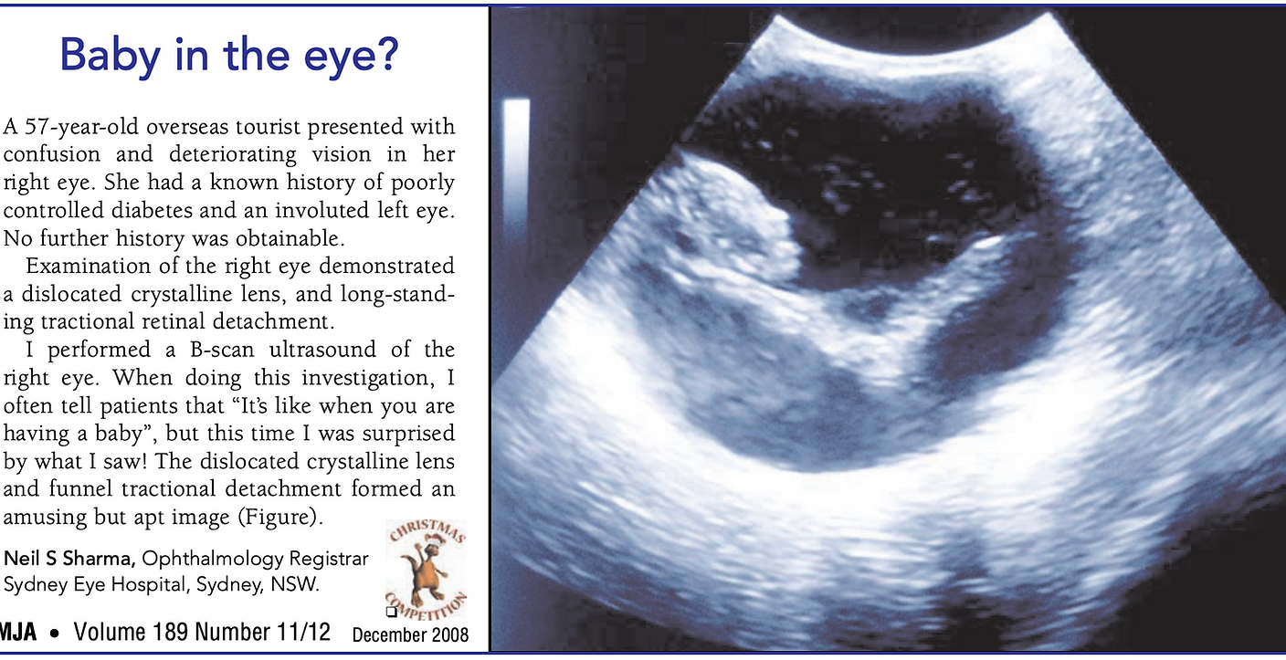
Ophthalmic ultrasound imaging remains an important investigation in modern retinal and retinal practice, particularly when direct visualisation of intraocular structures is limited by media opacity or complex posterior segment disease.
At Eye and Retina Specialists, B-scan ultrasonography is used in assessment of retinal detachment, vitreous haemorrhage, ocular tumours and other vitreoretinal pathology where conventional retinal imaging alone is insufficient.
Ultrasound imaging is particularly valuable in patients with very dense cataracts where direct examination of the retina is not possible clinically. In these situations, ultrasound assessment assists in excluding retinal detachment, intraocular masses and other significant posterior segment pathology prior to cataract surgery. This allows important posterior segment abnormalities to be identified despite severe media opacity, and assists in surgical planning and patient counselling prior to cataract intervention.
As ophthalmic ultrasound imaging is performed dynamically in real time, it allows assessment of movement and spatial relationships of intraocular structures during examination. This provides additional clinical information regarding vitreoretinal traction, choroidal detachment and posterior segment anatomy that may not always be apparent on static imaging alone.
.