
Clinical Education Articles for Referrers
In addition to peer-reviewed publications within ophthalmology literature, Dr Neil Sharma and colleagues have contributed educational articles for primary care clinicians, including publications in the Australian Journal of General Practice (AJGP), the official journal of the Royal Australian College of General Practitioners, ANZ Journal of Surgery and Australian Journal of Rural Health.
These publications examine clinically relevant ophthalmic presentations encountered in general practice and primary care, including urgent neuro-ophthalmic conditions, retinal disease and potentially sight-threatening or life-threatening presentations requiring timely specialist assessment. The articles additionally discuss important clinical examination findings, differential diagnoses, initial investigation and management strategies, and practical referral considerations relevant to primary care practice.
Ophthalmic Emergencies and Neuro-Ophthalmology
These publications examine urgent ophthalmic and neuro-ophthalmic presentations encountered in primary care and emergency settings, including potentially sight-threatening and life-threatening conditions requiring prompt recognition, investigation and specialist referral.
Sudden Loss of Vision
Australian Family Physician (AFP), 2013
Key topics: retinal artery occlusion, retinal detachment, vitreous haemorrhage, giant cell arteritis, ophthalmic emergencies
Article Summary: This publication reviews the differential diagnosis and urgent assessment of sudden visual loss, a presentation frequently encountered in both general practice and emergency medicine. The article discusses important causes of acute visual loss including retinal detachment, vitreous haemorrhage, retinal vascular occlusion, optic neuropathy and giant cell arteritis.
The publication emphasises the importance of careful history-taking and targeted examination in differentiating ocular from neurological causes of visual loss. Important clinical warning signs are reviewed, including visual field defects, photopsia, floaters, afferent pupillary defects and symptoms suggestive of giant cell arteritis such as headache, scalp tenderness and jaw claudication.
The article additionally outlines practical referral considerations and highlights the need for urgent ophthalmic assessment in patients with potentially reversible causes of visual loss.

Bilateral Optic Disc Swelling in a Man Aged 32 Years
Australian Journal of General Practice (AJGP), 2023
Key topics: optic disc swelling, neurosyphilis, neuro-ophthalmology, papilloedema, infectious disease
Article Summary: This publication reviews the differential diagnosis and investigation of bilateral optic disc swelling in a young man presenting with progressive blurred vision, with particular emphasis on the ophthalmic manifestations of neurosyphilis.
The article describes the presentation of a 32-year-old man with progressive bilateral visual disturbance associated with severe bilateral optic disc swelling, retinal nerve fibre layer haemorrhages and retinal folds. The publication outlines the broad differential diagnosis of bilateral optic disc swelling, including raised intracranial pressure, inflammatory optic neuropathies, vascular disease, intracranial space-occupying lesions and infectious conditions such as syphilis and tuberculosis.
The article emphasises the importance of urgent neuroimaging in patients presenting with bilateral optic disc swelling and reviews the stages, investigation and treatment of neurosyphilis, together with broader implications for ophthalmic and primary care practice.

New-Onset Ptosis Initially Diagnosed as Conjunctivitis
Australian Family Physician (AFP), 2015
Key topics: third nerve palsy, intracranial aneurysm, neuro-ophthalmology, ptosis, diplopia
Article Summary: This publication examines the distinction between neurogenic ptosis and inflammatory eyelid swelling, highlighting how potentially life-threatening neurological pathology may initially be mistaken for common ocular surface disease.
The article describes the case of a 66-year-old man initially treated for presumed conjunctivitis who subsequently developed worsening periocular pain, diplopia and near-complete ptosis. Ophthalmic examination demonstrated a pupil-involving third cranial nerve palsy with associated ocular motility abnormalities. Urgent neuroimaging subsequently revealed a posterior communicating artery aneurysm requiring neurosurgical intervention.
The publication reviews the differential diagnosis of third nerve palsy and discusses the importance of pupillary assessment, ocular motility examination and urgent referral in patients presenting with ptosis, diplopia or anisocoria.

A Painful, Red Eye
Australian Family Physician (AFP), 2009
Key topics: herpes simplex keratitis, anterior uveitis, contact lens complications, corneal ulceration
Article Summary: This publication reviews the assessment of the painful red eye and outlines important clinical features that may assist primary care clinicians in distinguishing benign ocular surface irritation from potentially sight-threatening disease.
The article discusses important differential diagnoses including microbial keratitis, herpes simplex keratitis, anterior uveitis, acute angle closure glaucoma and scleritis. Particular emphasis is placed on warning signs requiring urgent ophthalmic referral, including photophobia, reduced vision, corneal opacity and contact lens-associated infection.
The publication additionally discusses the potential risks associated with inappropriate corticosteroid use in undiagnosed corneal disease and reinforces the importance of timely specialist assessment in patients with severe ocular pain or visual disturbance.

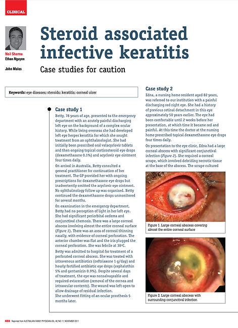
Steroid Associated Infective Keratitis
Australian Family Physician (AFP), 2011
Key topics: corticosteroids, microbial keratitis, herpes simplex keratitis, corneal ulceration
Article Summary: This publication examines the complications associated with topical corticosteroid use in patients with undiagnosed infective keratitis.
The article reviews the clinical course of steroid-associated corneal infection and discusses important causes including herpes simplex keratitis and bacterial keratitis. Particular emphasis is placed on the risk of delayed diagnosis, progressive corneal thinning and irreversible visual loss associated with inappropriate corticosteroid treatment.
The publication additionally discusses practical prescribing considerations and reinforces the importance of careful ophthalmic examination prior to commencing topical corticosteroid therapy in patients with red eye presentations.
Acute Infective Endophthalmitis
Australian Family Physician (AFP), 2012
Key topics: postoperative infection, endophthalmitis, ophthalmic emergency, cataract surgery complications
Article Summary: This publication reviews acute infective endophthalmitis, a severe intraocular infection associated with rapid visual deterioration and potentially profound visual loss.
The article discusses common clinical presentations following cataract surgery or intravitreal procedures, including ocular pain, hypopyon, reduced vision and severe intraocular inflammation. Important risk factors, microbiological considerations and management principles are reviewed.
The publication emphasises the importance of urgent ophthalmic referral and early treatment, including intravitreal antibiotics and vitreous sampling, in order to optimise visual outcomes.

The Length of Superficial Temporal Artery Biopsies
Australian and New Zealand (ANZ) Journal of Surgery, 2007
Key topics: giant cell arteritis, temporal artery biopsy, vasculitis, diagnostic accurac
Article Summary: This comparative study evaluated temporal artery biopsy specimen lengths obtained at both a tertiary referral centre and a regional hospital in patients undergoing investigation for suspected giant cell arteritis.
The publication demonstrated that biopsy specimens measuring 20 mm or greater were significantly more likely to demonstrate histopathological features of giant cell arteritis than shorter specimens. The study highlighted the importance of obtaining adequately sized biopsy specimens in order to reduce false-negative results associated with skip lesions and segmental arterial involvement.
The article additionally discusses practical diagnostic considerations relevant to clinicians involved in the assessment and management of giant cell arteritis, an important ophthalmic emergency associated with irreversible visual loss if not recognised and treated promptly.

Increase in the Length of Superficial Temporal Artery Biopsy Over 14 Years
Clinical and Experimental Ophthalmology, 2016
Key topics: corticosteroids, giant cell arteritis, temporal artery biopsy, ophthalmic vasculitis, quality improvement
Article Summary: This study reviewed temporal artery biopsy practice patterns over a 14-year period at a tertiary referral centre and evaluated whether biopsy specimen lengths had improved following earlier recommendations regarding optimal specimen size in suspected giant cell arteritis.
The publication demonstrated a significant increase in average biopsy specimen length over time compared with earlier institutional audit data. Despite this improvement, giant cell arteritis positivity rates remained relatively stable.
The article reinforces the importance of specimen adequacy and careful surgical technique in the investigation of giant cell arteritis and highlights the multidisciplinary nature of management involving ophthalmologists, physicians and primary care clinicians.

Retinal Disease and Medical Ophthalmology
These publications examine retinal disease, systemic associations and evidence based medical ophthalmology relevant to both specialist and primary care practice.
The Use of Fenofibrate in the Management of Patients with Diabetic Retinopathy
Australian Family Physician (AFP), 2015
Key topics: diabetic retinopathy, fenofibrate, diabetes, retinal vascular disease
Article Summary: This publication reviews the role of fenofibrate in the management of diabetic retinopathy and discusses emerging evidence supporting its use in reducing progression of retinal vascular disease in selected patients with diabetes.
The article reviews important findings from the FIELD and ACCORD-Eye studies and discusses the potential mechanisms by which fenofibrate may influence retinal vascular permeability, inflammation and microvascular disease progression.
The publication additionally discusses multidisciplinary management of diabetic retinopathy and reinforces the importance of systemic risk factor optimisation, including glycaemic control, blood pressure management and regular ophthalmic assessment.

Unilateral Cataract: A Potentially Fatal Prognosis
Australian Journal of General Practice (AJGP), 2018
Key topics: uveal melanoma, cataract, ocular oncology, retinal diseas
Article Summary: This publication examines the diagnostic challenges associated with unilateral cataract and emphasises the importance of considering posterior segment pathology in patients presenting with asymmetric or atypical visual symptoms.
The article describes the case of a 67-year-old man referred for cataract assessment after experiencing progressive unilateral visual blurring. Dilated fundus examination revealed a large ciliochoroidal melanoma initially obscured by lens opacity.
The publication reviews important clinical features suggestive of posterior segment disease, including asymmetry of vision, abnormalities of the red reflex and associated retinal findings, and reinforces the importance of careful ophthalmic examination in patients presenting with atypical cataract symptoms.

Rural and Indigenous Eye Health
These publications discuss ophthalmic disease encountered in rural and remote clinical settings, including Indigenous eye health, ocular trauma and preventable visual impairment.
Dislocated Crystalline Lens in an Aboriginal Patient
Australian Journal of Rural Health (AJRH), 2012
Key topics: traumatic cataract, lens dislocation, retinal detachment, Indigenous eye health, rural ophthalmology
Article Summary: This publication describes the assessment of a 41-year-old Aboriginal patient from a remote Northern Territory community presenting with profound bilateral visual impairment. Examination revealed complete dislocation of the crystalline lens into the vitreous cavity in one eye, associated with chronic retinal detachment and traumatic mydriasis, together with a dense cataract in the fellow eye.
The article discusses the importance of considering traumatic and non-age-related causes of cataract in younger patients, particularly in rural and remote clinical settings where access to ophthalmic assessment may be limited. Clinical signs suggestive of previous ocular trauma are reviewed, including traumatic mydriasis, zonular instability and retinal detachment.
The publication additionally discusses the broader issue of preventable visual impairment within Indigenous Australian communities and highlights the importance of timely recognition, detailed clinical history-taking and appropriate referral pathways in rural and primary care practice.


Education and Referrer Collaboration
At Eye and Retina Specialists, we value collaborative relationships with general practitioners, optometrists, ophthalmologists and other medical specialists involved in the care of our patients. Our doctors are involved in training, teaching and research and welcome contact by colleagues wishing to collaborate. We host educations seminars for optometrists, general practitioners and other health professionals.

Email us at reception@eyeandretina.com.au to be added to our invitation mailing list for future education events